Home » Without Label » Loculated Pleural Effusion : Loculated pleural effusion | Radiology Case | Radiopaedia.org / In general, pleural effusions can be divided into transudates (caused by fluid leaking from blood vessels) and exudates (where fluid leaks from inflammation of the pleura and lung).
Loculated Pleural Effusion : Loculated pleural effusion | Radiology Case | Radiopaedia.org / In general, pleural effusions can be divided into transudates (caused by fluid leaking from blood vessels) and exudates (where fluid leaks from inflammation of the pleura and lung).
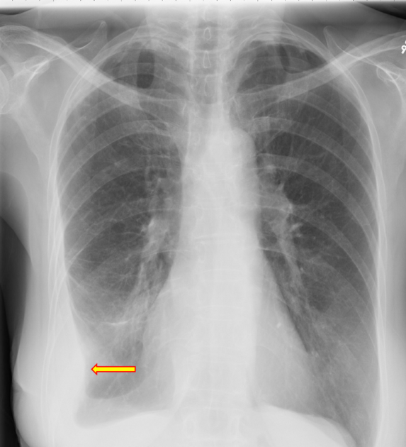
Loculated Pleural Effusion : Loculated pleural effusion | Radiology Case | Radiopaedia.org / In general, pleural effusions can be divided into transudates (caused by fluid leaking from blood vessels) and exudates (where fluid leaks from inflammation of the pleura and lung).. If it is clear that there are multiple loculations then it is wise to avoid delay and proceed directly to this procedure. Tell a friend about us, add a link to this page, or visit the webmaster's page for free fun content. A pleural effusion is due to the manifestations of another illness.; Left pleural effusion with high density material at the posterior costophrenic angle. Pleural fluid is seen extending to the right oblique fissure.
Streptokinase appears to improve the resolution of loculated pleural effusions when chest tube drainage fails to achieve symptomatic relief. Pleural effusion is a rare complication and is generally similar to that seen in the rickettsial illnesses. Most effusions start like this and can be easily missed. Tube thoracostomy has variable success in the treatment of complex pleural effusions, with In chf effusions are bilateral and more on right.
A) Loculated pleural effusion. A complex pleural effusion ... from www.researchgate.net Pleural effusions are very common, and physicians of allspecialties encounter them. Icu patients cannot sit up and the effusion layers posteriorly. Most pleural effusions, whether free flowing or loculated, are hypoechoic with a sharp echogenic line that delineates the visceral pleura and lung. Pleural effusion is when fluid fills this gap and separates the lungs from the chest wall. In chf effusions are bilateral and more on right. The pleura are thin membranes that line the lungs and the inside of the chest cavity and act to lubricate and facilitate breathing. Loculated pleural effusion the pleura is a thin membrane between the lungs and chest wall that lubricates these surfaces and allows movement of the lungs while breathing. Normally, a small amount of fluid is present in the pleura.
Pleural effusion is when fluid fills this gap and separates the lungs from the chest wall.
Icu patients cannot sit up and the effusion layers posteriorly. Encysted pleural fluid is visualized between the right upper and middle lobe (s). Pleural effusion that is confined to one or more fixed pockets in the pleural space. Tube thoracostomy has variable success in the treatment of complex pleural effusions, with Surgical thoracostomy tube placement and radiologically guided catheter drainage are standard therapy for loculated pleural fluid collections. Pleural effusions are very common, and physicians of allspecialties encounter them. Most effusions start like this and can be easily missed. The pleural space is normally filled with ~5 to 10 ml of serous fluid, which is secreted mainly from the parietal pleura at a rate of 0.01 ml/kg/h and absorbed through the lymphatics. Most malignant effusions can be controlled by thoracentesis and/or closed thoracostomy tube drainage and sclerosis of the pleural cavity. The lack of specificity is mainly due to the limitations of the imaging modality. What are the different appearances of pleural effusion? In chf effusions are bilateral and more on right. There are no established guidelines to facilitate management of nmpes and most management strategies rely on expert experience and data derived from patients with malignancy.
Nonmalignant pleural effusions (nmpes) have a wide variety of etiologies ( table 1 and table 2 and table 3) and cause significant morbidity and mortality 2,3 . Most effusions start like this and can be easily missed. (vats) with lysis of adhesions is also a viable option for loculated effusions. Complex septated, complex nonseptated, or homogeneously echogenic effusions are always exudates (fig. A rationaldiagnostic workup, emphasizing the most commoncauses, will reveal the etiology in most cases.
Aspiration of loculated pleural effusion - YouTube from i.ytimg.com (vats) with lysis of adhesions is also a viable option for loculated effusions. Treatment may fail if the catheter is not placed optimally within the loculation or if the fluid is hemorrhagic or fibrinous. Pleural fluid is seen extending to the right oblique fissure. Of loculated pleural effusions* jeffreys. Loculated malignant effusions however, are inherently resistant to the usual approaches because of nonexpanding underlying lung. Surgical thoracostomy tube placement and radiologically guided catheter drainage are standard therapy for loculated pleural fluid collections. Loculated pleural effusion the pleura is a thin membrane between the lungs and chest wall that lubricates these surfaces and allows movement of the lungs while breathing. Tell a friend about us, add a link to this page, or visit the webmaster's page for free fun content.
Pleural effusions describe fluid between the two layer of tissue (pleura) that cover the lung and the lining of the chest wall.
1 pleural effusion is defined as abnormal fluid collection in the pleural space. Most pleural effusions, whether free flowing or loculated, are hypoechoic with a sharp echogenic line that delineates the visceral pleura and lung. A pleural effusion representsthe disruption of the normal mechanisms of formationand drainage of fluid from the pleural space. Pleural effusion that is confined to one or more fixed pockets in the pleural space. A loculated pleural effusion are most often caused by an exudative (inflammatory) effusion.
Chest Radiograph from cdemcurriculum.files.wordpress.com Streptokinase appears to improve the resolution of loculated pleural effusions when chest tube drainage fails to achieve symptomatic relief. Of loculated pleural effusions* jeffreys. All patients require medical management with antibiotics. 1 pleural effusion is defined as abnormal fluid collection in the pleural space. The lack of specificity is mainly due to the limitations of the imaging modality. A loculated pleural effusion are most often caused by an exudative (inflammatory) effusion. Tube thoracostomy has variable success in the treatment of complex pleural effusions, with Pleural effusion that is confined to one or more fixed pockets in the pleural space.
What are the different appearances of pleural effusion?